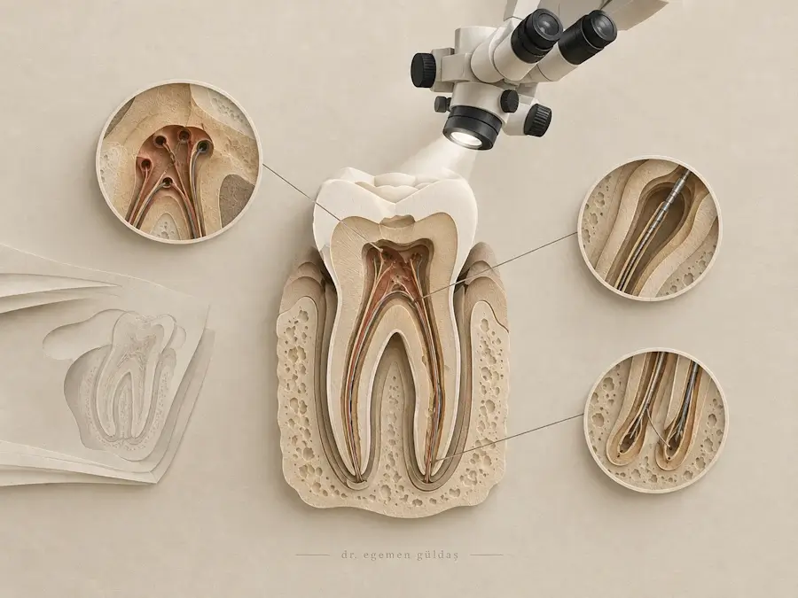
Microscopic endodontics uses a dental operating microscope to provide magnified vision and focused illumination during endodontic diagnosis and treatment. This approach is particularly useful when working with narrow root canals, complex internal anatomy, calcified canal openings or complications from previous dental procedures.
Dr. Egemen Güldaş, DDS, PhD, is an endodontist in Antalya who evaluates conditions affecting the dental pulp and root canal system. The use of magnification can support the identification of anatomical details that may be difficult to see with unaided vision, helping guide treatment decisions in selected cases.
Microscopic techniques may be used during initial root canal treatment, retreatment and certain endodontic procedures. Whether a microscope is indicated depends on the condition of the tooth, the complexity of the case and the treatment objectives.
For patients living in Antalya or travelling to Türkiye for dental care, an endodontic assessment helps determine the appropriate procedure and whether magnification may be beneficial.
What Is Microscopic Endodontics?
Microscopic endodontics refers to endodontic procedures performed with the aid of a dental operating microscope. The microscope combines magnification with directed illumination, allowing the clinician to examine small structures within the tooth in greater detail.
The pulp chamber and root canal system can contain narrow openings, additional canals, calcifications and anatomical variations. These features may complicate treatment, particularly when a tooth has undergone previous procedures or has extensive restorations.
Magnified visualisation can assist in locating canal openings, examining the pulp chamber floor and identifying certain structural abnormalities. It can also support the controlled use of fine instruments during procedures that require access to restricted areas.
A microscope is an aid to clinical diagnosis and treatment rather than a separate treatment for a specific disease. The underlying condition still determines which endodontic procedure is required.
When Is Microscopic Endodontics Recommended?
Microscopic techniques are especially relevant when the internal anatomy of a tooth is difficult to assess or when a procedure requires detailed visual control.
Complex Root Canal Anatomy
Some teeth have additional canals, narrow canal openings or curved root canal systems. These variations can make it more difficult to identify and access every canal.
Magnification may help the endodontist examine the pulp chamber floor and locate canal openings that are difficult to distinguish during conventional visual inspection.
Calcified Root Canals
Calcification can narrow or partially obstruct the root canal system, making access more challenging. This may occur with ageing, previous dental treatment, longstanding irritation or dental trauma.
Microscopic visualisation can help identify anatomical landmarks and guide the careful removal of dentine when access to a calcified canal is clinically appropriate. The extent of calcification and the position of the canal influence whether it can be safely located and treated.
Root Canal Retreatment
A tooth that has previously undergone root canal treatment may require further treatment if infection persists or returns. During retreatment, magnification can assist in identifying missed canals, examining existing access cavities and managing certain obstructions.
The use of a microscope does not guarantee that every obstruction can be removed or that every previously treated tooth can be preserved. The feasibility of retreatment depends on the individual findings.
Separated Instruments
An endodontic instrument can occasionally separate inside a root canal during treatment. Whether the fragment needs to be removed, bypassed or left in place depends on its position, the condition of the canal and the risks associated with retrieval.
Magnification can improve visibility when assessing the fragment and performing a removal procedure. The safest approach is determined by the potential benefit of retrieval compared with the risk of weakening or damaging the root.
Suspected Cracks or Structural Defects
Small cracks, perforations and other defects can be difficult to identify during a routine examination. A microscope may help the clinician inspect accessible areas of the tooth and assess certain structural abnormalities.
However, not every crack is visible, and microscopic examination alone cannot establish the full extent or prognosis of every fracture. Additional diagnostic methods may be needed.
How Is Microscopic Endodontic Treatment Performed?
The exact procedure depends on the diagnosis. The microscope is incorporated into the relevant stages of treatment rather than replacing standard endodontic principles.
1. Clinical Examination and Treatment Planning
The tooth is examined, symptoms and previous dental treatment are reviewed, and appropriate diagnostic tests are performed. Dental radiographs help assess the roots and surrounding tissues.
If the case involves unusual anatomy, calcification, persistent infection or a suspected complication, additional imaging may be considered when clinically justified.
The findings determine whether microscopic techniques are likely to contribute to the planned procedure.
2. Magnified Examination of the Tooth
The dental operating microscope provides adjustable magnification and focused illumination. The clinician can examine the access cavity, pulp chamber floor and visible canal openings in greater detail.
This helps guide decisions about where to continue treatment and which areas require closer inspection.
3. Access to the Root Canal System
When root canal treatment is indicated, an opening is prepared to access the pulp chamber. Under magnification, the clinician can examine the chamber and identify the available canal openings.
The objective is to obtain appropriate access while preserving sound tooth structure and avoiding unnecessary removal of dentine.
4. Cleaning and Disinfection
Once the canals have been located and accessed, they are cleaned, shaped and disinfected using appropriate endodontic instruments and irrigation techniques.
The microscope supports visual control during access and selected technical procedures, but cleaning and disinfection also depend on instrument selection, irrigation, canal anatomy and the overall treatment protocol.
5. Management of Specific Complications
In selected cases, magnification may assist with the management of calcified canals, separated instruments or certain perforations. These procedures require careful evaluation because attempts to gain access or remove an obstruction can create additional risks.
The clinician determines whether intervention is appropriate and explains the available options when the outcome is uncertain.
6. Root Canal Filling and Restoration
After the canal system has been adequately prepared, it is filled and sealed when indicated. The tooth is then restored according to the amount of remaining structure, its location and its functional requirements.
Follow-up may be recommended to assess symptoms and healing around the root.
What Are the Benefits of Using a Dental Operating Microscope?
The principal benefit of microscopic endodontics is improved visualisation of small structures during selected procedures. The clinical value depends on the condition being treated and the clinician’s ability to interpret the findings.
Improved Visualisation of Canal Openings
Magnification and focused illumination can help distinguish small canal openings and anatomical details within the pulp chamber. This may be particularly useful when additional canals are suspected.
Greater Control During Complex Procedures
Detailed visualisation can support controlled instrument use when managing calcifications, obstructions and certain procedural complications.
Support for Conservative Treatment
When anatomy can be identified more clearly, the clinician may be able to target access and remove dentine selectively rather than relying on broader exploration. The amount of tooth structure that can safely be preserved still depends on the anatomy and clinical requirements.
Assistance in Evaluating Treatment Complications
A microscope can help examine accessible areas when investigating missed canals, perforations or certain structural defects. The findings contribute to decisions about whether further treatment is feasible.
Magnification does not eliminate procedural risks, replace diagnostic imaging or guarantee a favourable outcome. It is one component of a treatment approach that also depends on clinical judgement, technique and the condition of the tooth.
Microscopic Endodontics and the Role of an Endodontist
Endodontic procedures often involve structures that are difficult to examine directly. An endodontist’s training focuses on diagnosing and treating diseases of the dental pulp and tissues surrounding the roots.
Dr. Egemen Güldaş, DDS, PhD, evaluates endodontic conditions according to the individual tooth, its internal anatomy and the clinical problem requiring treatment.
The decision to use a dental operating microscope is based on whether additional visual detail is likely to assist diagnosis or treatment. Depending on the case, microscopic techniques may be incorporated into initial root canal treatment, retreatment or the management of selected complications.
The objective is to select an appropriate procedure based on clinical findings rather than using the same technique for every patient.
Microscopic Endodontics for International Patients in Antalya
Patients who live in Antalya, visit the city on holiday or travel to Türkiye for dental treatment may need to coordinate appointments with their work or travel arrangements.
The treatment schedule depends on the underlying condition, the complexity of the procedure and whether additional restorative care is required. Some procedures can be completed in one appointment, while others require several visits or follow-up after treatment.
Patients with previous root canal treatment, dental trauma or a known endodontic complication may bring earlier radiographs and treatment records to support the initial assessment. These documents can provide useful background information but do not replace a clinical examination.
Before treatment begins, the proposed plan should clarify the procedure required, the likely appointment schedule and any need for a permanent restoration or subsequent review.
Frequently Asked Questions About Microscopic Endodontics
Is microscopic endodontics different from ordinary root canal treatment?
The underlying principles of root canal treatment remain the same. Microscopic endodontics uses magnification and focused illumination to improve visualisation during selected stages of diagnosis and treatment.
Does every root canal treatment require a dental microscope?
No. The need for magnification depends on the tooth’s anatomy, the clinical findings and the complexity of the procedure. Some cases can be managed without a microscope, while others may benefit from its use.
Can a dental microscope help locate a missed root canal?
Magnification can improve the visibility of canal openings and may help identify a canal that was not located during an earlier procedure. However, success depends on the canal’s position, calcification, accessibility and the overall anatomy of the tooth.
Can microscopic endodontics save a tooth that has failed root canal treatment?
Microscopic techniques may assist with retreatment when a correctable cause of persistent disease can be identified. Whether the tooth can be retained depends on factors such as root condition, structural damage, fracture risk and the feasibility of restoring it.
Is microscopic endodontic treatment painful?
The level of discomfort depends on the procedure and the condition of the tooth. Local anaesthesia is used when appropriate. Some tenderness may occur afterwards, particularly when inflammation or infection was present before treatment.
Can international patients receive microscopic endodontic treatment during a short stay in Antalya?
Treatment may be possible during a short visit, but the schedule depends on the diagnosis and clinical requirements. Some cases need additional appointments, definitive restoration or follow-up. An assessment is necessary before a realistic treatment schedule can be established.
Microscopic Endodontic Assessment in Antalya
If you have been advised to undergo complex root canal treatment, have a calcified canal or require further assessment after previous endodontic treatment, a specialist examination can help determine the available options.
Dr. Egemen Güldaş, DDS, PhD, provides endodontic assessment and treatment in Antalya. The use of microscopic techniques is determined by the clinical findings and whether magnification can assist the planned procedure.